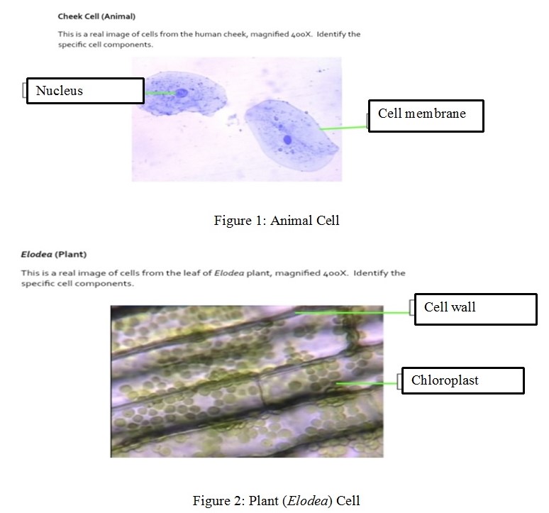
Cheek Cell (animal) – the marked cell components are the nucleus and cell membrane. Elodea (plant) – the marked cell components are the cell wall and chloroplast that appear as the green dots.

A compound microscope’s total magnifying power is determined by multiplying the ocular lens’ magnifying power with the objective lens’ magnifying power. As such, I would use the 10X objective lens together with the 10X ocular lens to achieve the 100X magnification.
The main difference between the fungi (mold or yeast cells) and the Elodea cell is that fungal cells are not photosynthetic because they lack photosynthetic pigments or chloroplasts. Engelkirk et al. (2020) also point out that the fungal cells also have cell walls made of chitin, while plant cells (such as Elodea) have cell walls composed of cellulose. The main difference between the mold or yeast cells and the cheek cell is that the fungal cells have cell walls while the cheek cells do not have the cell wall.
The two different shapes I observed on the bacteria cells are the spiral and rod shapes. Typing the letter “O” at scanning power allowed the student to focus on the compound microscope and view the image while positioned at the center of the field. However, increasing the magnification using the higher-powered objective lens causes the letter “O” to disappear because the field of view becomes narrower with increasing magnification power. The letter did not disappear completely from the field, but the focus changed, and the student could not see it without replacing the first objective lens.
Reference
Engelkirk, P. G., Duben-Engelkirk, J., & Fader, R. C. (2020). Burton’s microbiology for the health sciences, enhanced edition. Jones & Bartlett Learning.
